Creating Menus
in CSS Css3Menu.com
The retinal
examination:
Indirect ophthalmoscopy - visualization of the retina with a head-lamp
and handheld lens to obtain a panoramic, three-dimensional (3-D) view
of the macula, optic nerve and retinal periphery. The indirect
ophthalmoscope is the instrument of choice in viewing the peripheral
retina for retinal tears and detachment. Despite its simplicity
of design, it requires years of practice to gain facility as the viewed
magnified image is inverted up-side-down and reversed left to right.
And
may also
include:
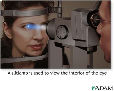
Biomicroscopy - visualization of the retina with a slit lamp and
handheld or contact lens provides a 3-D view with increased
magnification.
Fundus photography - provides a high power two-dimensional view and
record of the posterior retina including the disc and macula.
Fluorescein angiography - fundus photography performed with the
simultaneous injection of fluorescein dye into the systemic circulation
highlights vascular disease and disorders in the retina, RPE and
choroid. The angiogram helps identify damaged blood
vessels, the quality of blood flow through the retina, and neovascular
elements - in a flat plane of two dimensions.
Ocular coherence tomography - a retinal study with computerized low
intensity laser scanning of the retina provides microscopic detail of
the retinal structure and orientation. Three-dimensional OCT
gives precise analysis of retinal thickness and contour through the
macular region in a three second non-contact sweep. OCT helps
identify macular edema, macular holes, choroidal neovascularization and
vitreo-retinal interface disturbances.
Indocyanine green (ICG) angiography - fundus photography with ICG
highlights occult choroidal neovascularization with low-grade leakage.
Automated visual field testing - tests the sensitivity of central and
peripheral visual perception – often to help assess the integrity of
the optic nerve.
Electrophysiology - special testing of the magnitude of electrical
polarization and discharge within the retina under varying levels of
illumination, is especially helpful to assess abnormalities in rod and
cone function. This testing helps identify and categorize
retinitis pigmentosa, cone-rod dystrophies, night blindness, retinal
dystrophies and ischemia.